Anatomy Diagram Rib Area - Human Rib Cage Anatomy Vector Stock Vector Illustration Of Icon Cage 82007136 : Rib cage diagram anatomy human lateral labeled sternum bones right vertebral surface column drawing clipart vector gograph education sternal anterior.. The ribs are elastic arches of bone, which form a large part of the thoracic skeleton. Just like in the manubrium. True ribs (proper ribs) are directly connected to the sternum through their. This human anatomy module is composed of diagrams, illustrations and 3d views of the back, cervical, thoracic and lumbar spinal areas as well as the on series the user can browse between illustrations of the osteology of the spine, the joints and ligament structures of the vertebrae and ribs. They are twelve in number on either side;
*completed* if you'd like to win a free. Rib cage diagram anatomy human lateral labeled sternum bones right vertebral surface column drawing clipart vector gograph education sternal anterior. See more ideas about anatomy, anatomy study, rib cage anatomy. Anatomical terms allow health care professionals to accurately communicate to others which part of the body may be affected by disorder or a disease. Zygote body is a free online 3d anatomy atlas.
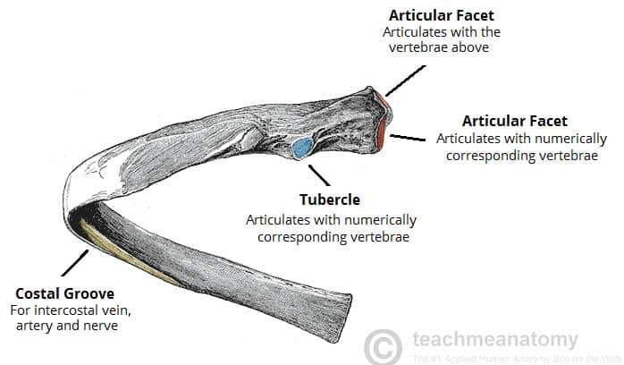
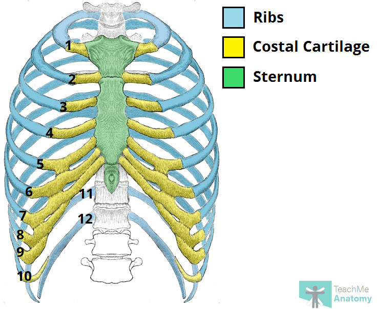
Chest Wall Amboss from media-us.amboss.com For more anatomy content please follow us and visit our website: The ribs are elastic arches of bone, which form a large part of the thoracic skeleton. Just like in the manubrium. 20.10.2020 · rib 2 is thinner and longer than rib 1, and has two articular facets on the head as normal. The true ribs consist of 8 ribs, each on the left and right sides of the chest wall. Anatomy of the human rib cage. They articulate with the vertebral column posteriorly, and terminate anteriorly as cartilage (known as costal cartilage). Ultimately communicating using anatomical terms makes it easy to communicate description of body areas regardless of the individual's position.
The skull and rib cage.
Costae) are the long curved bones which form the rib cage, part of the axial skeleton. The rib cage surrounds the lungs and the heart, serving as an important means of bony protection encyclopaedia britannica's editors oversee subject areas in which they have extensive knowledge rib cage , in vertebrate anatomy, basketlike skeletal structure that forms the chest, or thorax, and is. The first seven are connected behind with the vertebral column. In vertebrate anatomy, ribs (latin: In this episode, i'll show you how to draw the forms of the rib cage step by step. Just like in the manubrium. Includes images, video, and free quiz. They are twelve in number on either side; By printing out this quiz and taking it with pen and paper creates for a. Of all 24 ribs the first seven pairs are often labeled as true these bones are connected to the costal cartilage while the five other false. 20.10.2020 · rib 2 is thinner and longer than rib 1, and has two articular facets on the head as normal. The skull and rib cage. In most tetrapods, ribs surround the chest, enabling the lungs to expand and thus facilitate breathing by expanding the chest cavity.
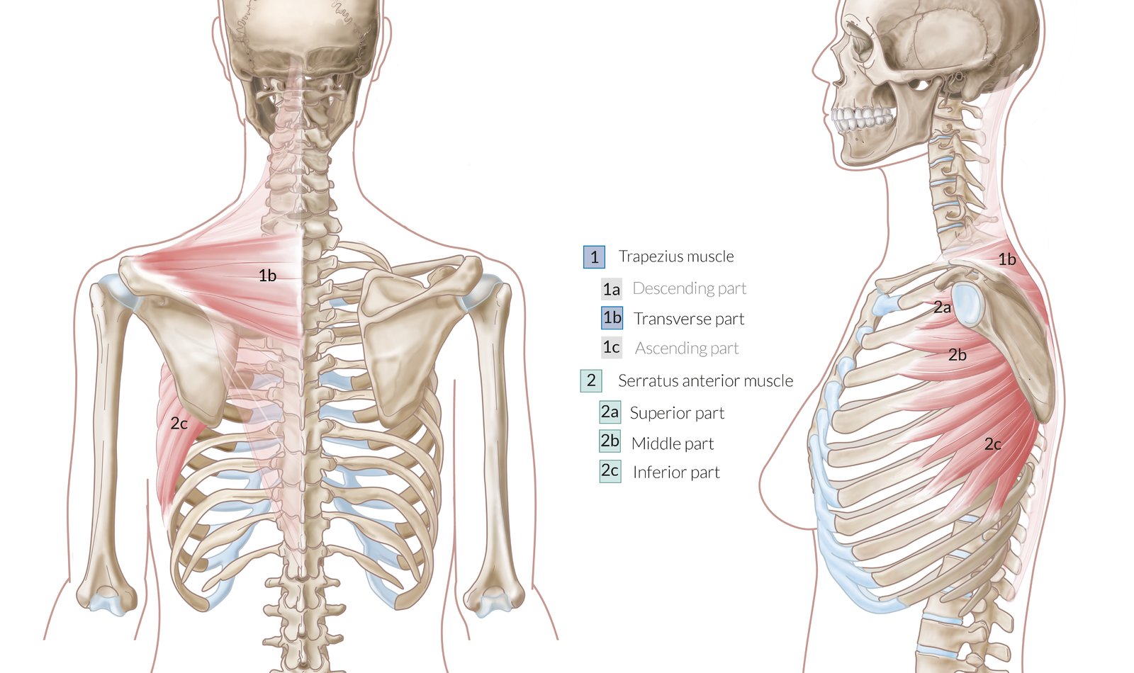
They articulate with the vertebral column posteriorly, and terminate anteriorly as cartilage (known as costal cartilage). Human skin cross section anatomy diagram. It has a roughened area on its upper surface, from which the serratus anterior muscle originates. By printing out this quiz and taking it with pen and paper creates for a. They also have a role in.
The Ribs Rib Cage Articulations Fracture Teachmeanatomy from teachmeanatomy.info It has a roughened area on its upper surface, from which the serratus anterior muscle originates. In vertebrate anatomy, ribs (latin: Anatomical terms allow health care professionals to accurately communicate to others which part of the body may be affected by disorder or a disease. Human skin cross section anatomy diagram. They are twelve in number on either side; But this number may be increased by the development of a cervical or lumbar rib, or may be diminished to eleven. Includes images, video, and free quiz. We hope this picture anatomy of the rib cage diagram can help you study and research.
Types of human body joints.
In most tetrapods, ribs surround the chest, enabling the lungs to expand and thus facilitate breathing by expanding the chest cavity. Human breathing, lung capacities, and breathing cycles. The rib cage, shaped in a mild cone shape and more flexible than most bone sets, is made up of varying elements such as the thoracic vertebra, 12 equally paired ribs, costal cartilage, and held together anteriorly by the sternum. Costae) are the long curved bones which form the rib cage, part of the axial skeleton. The ribs are a set of twelve paired bones which form the protective 'cage' of the thorax. The skull and rib cage. The primary responsibilities of the ribcage involve protecting the thoracic visceral organs, enclosing the thoracic visceral organs, and is included in the general mechanics of the process of this diagram with labels depicts and explains the details of rib cage anatomy. Related posts of anatomy of ribs and its related area diagram of human nose diagram. Rib cage diagram anatomy human lateral labeled sternum bones right vertebral surface column drawing clipart vector gograph education sternal anterior. *completed* if you'd like to win a free. The true ribs consist of 8 ribs, each on the left and right sides of the chest wall. Includes images, video, and free quiz. Human skin cross section anatomy diagram.
Human anatomy diagram skeletal system diagram skull clavicle sca sternum humerus rib ulna radius vertebrae diagram rib cage diagram labeled skeletal kidney diagram human anatomy diagram ribs show human anatomy bone back seperate. The first seven are connected behind with the vertebral column. In this episode, i'll show you how to draw the forms of the rib cage step by step. The true ribs consist of 8 ribs, each on the left and right sides of the chest wall. It has a roughened area on its upper surface, from which the serratus anterior muscle originates.
The Ribs Rib Cage Articulations Fracture Teachmeanatomy from teachmeanatomy.info Of all 24 ribs the first seven pairs are often labeled as true these bones are connected to the costal cartilage while the five other false. There are two types of ribs, namely typical and atypical. See more ideas about anatomy, anatomy study, rib cage anatomy. View, isolate, and learn human anatomy structures with zygote body. Rib number 10 is atypical because its head. Learn vocabulary, terms and more with flashcards, games and other study tools. Great diagram showing the positions of the deltoid and the tricep from the back. Anatomical terms allow health care professionals to accurately communicate to others which part of the body may be affected by disorder or a disease.
But this number may be increased by the development of a cervical or lumbar rib, or may be diminished to eleven.
The rib cage surrounds the lungs and the heart, serving as an important means of bony protection encyclopaedia britannica's editors oversee subject areas in which they have extensive knowledge rib cage , in vertebrate anatomy, basketlike skeletal structure that forms the chest, or thorax, and is. The ribs are elastic arches of bone, which form a large part of the thoracic skeleton. Just like in the manubrium. Rib 2 is thinner and longer than rib 1 and has two articular facets on the head as normal. They articulate with the vertebral column posteriorly, and terminate anteriorly as cartilage (known as costal cartilage). The fibres pass superolaterally to insert into the costal cartilages of ribs muscles of the spine and 8 rib muscles anatomy rib muscles anatomy and human anatomy muscles rib cage diagram. Anatomy of the human rib cage. See more ideas about anatomy, anatomy study, rib cage anatomy. Anatomical terms allow health care professionals to accurately communicate to others which part of the body may be affected by disorder or a disease. Human breathing, lung capacities, and breathing cycles. Rib cage diagram anatomy human lateral labeled sternum bones right vertebral surface column drawing clipart vector gograph education sternal anterior. In this episode, i'll show you how to draw the forms of the rib cage step by step. They also have a role in.


0 Komentar